Our website is made possible by displaying online advertisements to our visitors.
Please consider supporting us by disabling your ad blocker.
Golgi apparatus

| Cell biology | |
|---|---|
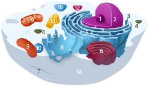
| Animal cell diagram | |
 Components of a typical animal cell:
|
The Golgi apparatus (/ˈɡɒldʒi/), also known as the Golgi complex, Golgi body, or simply the Golgi, is an organelle found in most eukaryotic cells.[1] Part of the endomembrane system in the cytoplasm, it packages proteins into membrane-bound vesicles inside the cell before the vesicles are sent to their destination. It resides at the intersection of the secretory, lysosomal, and endocytic pathways. It is of particular importance in processing proteins for secretion, containing a set of glycosylation enzymes that attach various sugar monomers to proteins as the proteins move through the apparatus.
The Golgi apparatus was identified in 1898 by the Italian biologist and pathologist Camillo Golgi.[2] The organelle was later named after him in the 1910s.[2]
- ^ Pavelk M, Mironov AA (2008). "Golgi apparatus inheritance". The Golgi Apparatus: State of the art 110 years after Camillo Golgi's discovery. Berlin: Springer. p. 580. doi:10.1007/978-3-211-76310-0_34. ISBN 978-3-211-76310-0.
- ^ a b Fabene PF, Bentivoglio M (October 1998). "1898-1998: Camillo Golgi and "the Golgi": one hundred years of terminological clones". Brain Research Bulletin. 47 (3): 195–8. doi:10.1016/S0361-9230(98)00079-3. PMID 9865849. S2CID 208785591.
Previous Page Next Page


