Our website is made possible by displaying online advertisements to our visitors.
Please consider supporting us by disabling your ad blocker.
Kidney stone disease
| Kidney stone disease | |
|---|---|
| Other names | Urolithiasis, kidney stone, renal calculus, nephrolith, kidney stone disease,[1] |
 | |
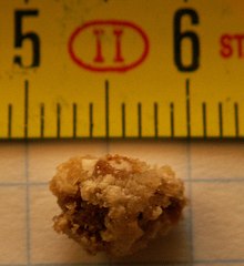
| A kidney stone, 8 millimeters (0.3 in) in diameter | |
| Specialty | Urology, nephrology |
| Symptoms | Severe pain in the lower back or abdomen, blood in the urine, vomiting, nausea[2] |
| Causes | Genetic and environmental factors[2] |
| Diagnostic method | Based on symptoms, urine testing, medical imaging[2] |
| Differential diagnosis | Abdominal aortic aneurysm, diverticulitis, appendicitis, pyelonephritis[3] |
| Prevention | Drinking fluids such that more than two liters of urine are produced per day[4] |
| Treatment | Pain medication, extracorporeal shock wave lithotripsy, ureteroscopy, percutaneous nephrolithotomy[2] |
| Frequency | 22.1 million (2015)[5] |
| Deaths | 16,100 (2015)[6] |
Kidney stone disease, also known as renal calculus disease, nephrolithiasis or urolithiasis, is a crystallopathy where a solid piece of material (renal calculus) develops in the urinary tract.[2] Renal calculi typically form in the kidney and leave the body in the urine stream.[2] A small calculus may pass without causing symptoms.[2] If a stone grows to more than 5 millimeters (0.2 inches), it can cause blockage of the ureter, resulting in sharp and severe pain in the lower back that often radiates downward to the groin (renal colic).[2][7] A calculus may also result in blood in the urine, vomiting, or painful urination.[2] About half of people who have had a renal calculus are likely to have another within ten years.[8]
Most calculi form by a combination of genetics and environmental factors.[2] Risk factors include high urine calcium levels, obesity, certain foods, some medications, calcium supplements, hyperparathyroidism, gout and not drinking enough fluids.[2][8] Calculi form in the kidney when minerals in urine are at high concentration.[2] The diagnosis is usually based on symptoms, urine testing, and medical imaging.[2] Blood tests may also be useful.[2] Calculi are typically classified by their location: nephrolithiasis (in the kidney), ureterolithiasis (in the ureter), cystolithiasis (in the bladder), or by what they are made of (calcium oxalate, uric acid, struvite, cystine).[2]
In those who have had renal calculi, drinking fluids is a way to prevent them. Drinking fluids such that more than two liters of urine are produced per day is recommended.[4] If fluid intake alone is not effective to prevent renal calculi, the medications thiazide diuretic, citrate, or allopurinol may be suggested.[4] Soft drinks containing phosphoric acid (typically colas) should be avoided.[4] When a calculus causes no symptoms, no treatment is needed.[2] For those with symptoms, pain control is usually the first measure, using medications such as nonsteroidal anti-inflammatory drugs or opioids.[7][9] Larger calculi may be helped to pass with the medication tamsulosin[10] or may require procedures such as extracorporeal shock wave lithotripsy, ureteroscopy, or percutaneous nephrolithotomy.[2]
Renal calculi have affected humans throughout history with a description of surgery to remove them dating from as early as 600 BC in ancient India by Sushruta.[1] Between 1% and 15% of people globally are affected by renal calculi at some point in their lives.[8][11] In 2015, 22.1 million cases occurred,[5] resulting in about 16,100 deaths.[6] They have become more common in the Western world since the 1970's.[8][12] Generally, more men are affected than women.[2][11] The prevalence and incidence of the disease rises worldwide and continues to be challenging for patients, physicians, and healthcare systems alike. In this context, epidemiological studies are striving to elucidate the worldwide changes in the patterns and the burden of the disease and identify modifiable risk factors that contribute to the development of renal calculi.[13]
- ^ a b Schulsinger DA (2014). Kidney Stone Disease: Say NO to Stones!. Springer. p. 27. ISBN 978-3-319-12105-5. Archived from the original on 8 September 2017.
- ^ a b c d e f g h i j k l m n o p q r "Kidney Stones in Adults". February 2013. Archived from the original on 11 May 2015. Retrieved 22 May 2015.
- ^ Knoll T, Pearle MS (2012). Clinical Management of Urolithiasis. Springer Science & Business Media. p. 21. ISBN 978-3-642-28732-9. Archived from the original on 8 September 2017.
- ^ a b c d Qaseem A, Dallas P, Forciea MA, et al. (November 2014). "Dietary and pharmacologic management to prevent recurrent nephrolithiasis in adults: a clinical practice guideline from the American College of Physicians". Annals of Internal Medicine. 161 (9): 659–67. doi:10.7326/M13-2908. PMID 25364887.
- ^ a b Vos T, Allen C, Arora M, et al. (GBD 2015 Disease and Injury Incidence and Prevalence Collaborators) (October 2016). "Global, regional, and national incidence, prevalence, and years lived with disability for 310 diseases and injuries, 1990-2015: a systematic analysis for the Global Burden of Disease Study 2015". Lancet. 388 (10053): 1545–1602. doi:10.1016/S0140-6736(16)31678-6. PMC 5055577. PMID 27733282.
- ^ a b Wang H, Naghavi M, Allen C, et al. (GBD 2015 Disease and Injury Incidence and Prevalence Collaborators) (October 2016). "Global, regional, and national life expectancy, all-cause mortality, and cause-specific mortality for 249 causes of death, 1980-2015: a systematic analysis for the Global Burden of Disease Study 2015". Lancet. 388 (10053): 1459–1544. doi:10.1016/s0140-6736(16)31012-1. PMC 5388903. PMID 27733281.
- ^ a b Miller NL, Lingeman JE (March 2007). "Management of kidney stones". BMJ. 334 (7591): 468–72. doi:10.1136/bmj.39113.480185.80. PMC 1808123. PMID 17332586.
- ^ a b c d Morgan MS, Pearle MS (March 2016). "Medical management of renal stones". BMJ. 352: i52. doi:10.1136/bmj.i52. PMID 26977089. S2CID 28313474.
- ^ Afshar K, Jafari S, Marks AJ, et al. (June 2015). "Nonsteroidal anti-inflammatory drugs (NSAIDs) and non-opioids for acute renal colic". The Cochrane Database of Systematic Reviews. 2015 (6): CD006027. doi:10.1002/14651858.CD006027.pub2. PMC 10981792. PMID 26120804.
- ^ Wang RC, Smith-Bindman R, Whitaker E, et al. (March 2017). "Effect of Tamsulosin on Stone Passage for Ureteral Stones: A Systematic Review and Meta-analysis". Annals of Emergency Medicine. 69 (3): 353–361.e3. doi:10.1016/j.annemergmed.2016.06.044. PMID 27616037.
- ^ a b Abufaraj M, Xu T, Cao C, et al. (November 2021). "Prevalence and Trends in Kidney Stone Among Adults in the USA: Analyses of National Health and Nutrition Examination Survey 2007-2018 Data". European Urology Focus. 7 (6): 1468–1475. doi:10.1016/j.euf.2020.08.011. PMID 32900675. S2CID 221572651.
- ^ Stamatelou KK, Francis ME, Jones CA, et al. (May 2003). "Time trends in reported prevalence of kidney stones in the United States: 1976–199411.See Editorial by Goldfarb, p. 1951". Kidney International. 63 (5): 1817–1823. doi:10.1046/j.1523-1755.2003.00917.x. PMID 12675858.
- ^ Stamatelou K, Goldfarb DS (January 2023). "Epidemiology of Kidney Stones". Healthcare. 11 (3): 424. doi:10.3390/healthcare11030424. ISSN 2227-9032. PMC 9914194. PMID 36766999.
 This article incorporates text from this source, which is available under the CC BY 4.0 license.
Previous Page Next Page


