Our website is made possible by displaying online advertisements to our visitors.
Please consider supporting us by disabling your ad blocker.
Limb development
| Development of the limbs | |
|---|---|
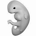
 Illustration of a human embryo at six weeks gestational age | |
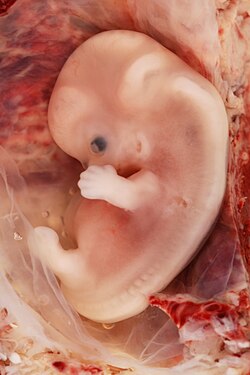
 9-week human fetus from ectopic pregnancy | |
| Anatomical terminology |
Limb development in vertebrates is an area of active research in both developmental and evolutionary biology, with much of the latter work focused on the transition from fin to limb.[1]
Limb formation begins in the morphogenetic limb field, as mesenchymal cells from the lateral plate mesoderm proliferate to the point that they cause the ectoderm above to bulge out, forming a limb bud. Fibroblast growth factor (FGF) induces the formation of an organizer at the end of the limb bud, called the apical ectodermal ridge (AER), which guides further development and controls cell death. Programmed cell death is necessary to eliminate webbing between digits.
The limb field is a region specified by expression of certain Hox genes, a subset of homeotic genes, and T-box transcription factors – Tbx5 for forelimb or wing development, and Tbx4 for leg or hindlimb development. Establishment of the forelimb field (but not hindlimb field) requires retinoic acid signaling in the developing trunk of the embryo from which the limb buds emerge.[2][3] Also, although excess retinoic acid can alter limb patterning by ectopically activating Shh or Meis1/Meis2 expression, genetic studies in mouse that eliminate retinoic acid synthesis have shown that RA is not required for limb patterning.[4]
The limb bud remains active throughout much of limb development as it stimulates the creation and positive feedback retention of two signaling regions: the AER and its subsequent creation of the zone of polarizing activity (ZPA) with the mesenchymal cells.[5] In addition to the dorsal-ventral axis created by the ectodermal expression of competitive Wnt7a and BMP signals respectively, these AER and ZPA signaling centers are crucial to the proper formation of a limb that is correctly oriented with its corresponding axial polarity in the developing organism.[6][7] Because these signaling systems reciprocally sustain each other's activity, limb development is essentially autonomous after these signaling regions have been established.[5]
- ^ Stewart, TA; Bhat, R; Newman, SA (2017). "The evolutionary origin of digit patterning". EvoDevo. 8: 21. doi:10.1186/s13227-017-0084-8. PMC 5697439. PMID 29201343.
- ^ Stratford T, Horton C, Maden M (1996). "Retinoic acid is required for the initiation of outgrowth in the chick limb bud". Curr Biol. 6 (9): 1124–33. Bibcode:1996CBio....6.1124S. doi:10.1016/S0960-9822(02)70679-9. PMID 8805369. S2CID 14662908.
- ^ Zhao X, Sirbu IO, Mic FA, et al. (June 2009). "Retinoic acid promotes limb induction through effects on body axis extension but is unnecessary for limb patterning". Curr. Biol. 19 (12): 1050–7. Bibcode:2009CBio...19.1050Z. doi:10.1016/j.cub.2009.04.059. PMC 2701469. PMID 19464179.
- ^ Cunningham, T.J.; Duester, G. (2015). "Mechanisms of retinoic acid signalling and its roles in organ and limb development". Nat. Rev. Mol. Cell Biol. 16 (2): 110–123. doi:10.1038/nrm3932. PMC 4636111. PMID 25560970.
- ^ a b Tickle, C (October 2015). "How the embryo makes a limb: determination, polarity and identity". Journal of Anatomy. 227 (4): 418–30. doi:10.1111/joa.12361. PMC 4580101. PMID 26249743.
- ^ Parr, BA; McMahon, AP (23 March 1995). "Dorsalizing signal Wnt-7a required for normal polarity of D-V and A-P axes of mouse limb". Nature. 374 (6520): 350–3. Bibcode:1995Natur.374..350P. doi:10.1038/374350a0. PMID 7885472. S2CID 4254409.
- ^ Pizette, S; Abate-Shen, C; Niswander, L (November 2001). "BMP controls proximodistal outgrowth, via induction of the apical ectodermal ridge, and dorsoventral patterning in the vertebrate limb". Development. 128 (22): 4463–74. doi:10.1242/dev.128.22.4463. PMID 11714672.
Previous Page Next Page


