Our website is made possible by displaying online advertisements to our visitors.
Please consider supporting us by disabling your ad blocker.
Oligodendrocyte
| Oligodendrocyte | |
|---|---|
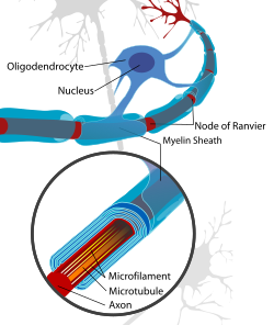
 Oligodendrocytes form the myelin insulation around the axons of neurons in the central nervous system | |
| Details | |
| Location | Central nervous system |
| Identifiers | |
| Latin | oligodendrocytus |
| MeSH | D009836 |
| TH | H2.00.06.2.00003, H2.00.06.2.01018 |
| FMA | 83665 54540, 83665 |
| Anatomical terms of microanatomy | |
Oligodendrocytes (from Greek 'cells with a few branches'), also known as oligodendroglia, are a type of neuroglia whose main function is to provide the myelin sheath to neuronal axons in the central nervous system (CNS). Myelination gives metabolic support to, and insulates the axons of most vertebrates.[1] A single oligodendrocyte can extend its processes to cover up to 40 axons, that can include multiple adjacent axons.[2] The myelin sheath is not continuous but is segmented along the axon's length at gaps known as the nodes of Ranvier. In the peripheral nervous system the myelination of axons is carried out by Schwann cells.[1]
Oligodendrocytes are found exclusively in the CNS, which comprises the brain and spinal cord. They are the most widespread cell lineage, including oligodendrocyte progenitor cells, pre-myelinating cells, and mature myelinating oligodendrocytes in the CNS white matter.[3] Non-myelinating oligodendrocytes are found in the grey matter surrounding and lying next to neuronal cell bodies. They are known as neuronal satellite cells, and their presence is not understood.[2]
It was once thought that oligodendrocytes were produced in the ventral neural tube, the embryonic precursor to the CNS. Studies have suggested that they originate from the ventral ventricular zone of the embryonic spinal cord, with some potential concentrations in the forebrain.[4] Oligodendrocytes are the last type of cell to be generated in the CNS.[5] Oligodendrocytes were discovered by Pío del Río Hortega.[6][7]
- ^ a b Carlson N (2010). Physiology of Behavior. Boston, MA: Allyn & Bacon. pp. 38–39. ISBN 978-0-205-66627-0.
- ^ a b Cite error: The named reference
Haines2018was invoked but never defined (see the help page). - ^ Seeker LA, Williams A (February 2022). "Oligodendroglia heterogeneity in the human central nervous system". Acta Neuropathol. 143 (2): 143–157. doi:10.1007/s00401-021-02390-4. PMC 8742806. PMID 34860266.
- ^ Richardson WD, Kessaris N, Pringle N (January 2006). "Oligodendrocyte wars". Nature Reviews. Neuroscience. 7 (1): 11–18. doi:10.1038/nrn1826. PMC 6328010. PMID 16371946.
- ^ Thomas JL, Spassky N, Perez Villegas EM, Olivier C, Cobos I, Goujet-Zalc C, et al. (February 2000). "Spatiotemporal development of oligodendrocytes in the embryonic brain". Journal of Neuroscience Research. 59 (4): 471–476. doi:10.1002/(SICI)1097-4547(20000215)59:4<471::AID-JNR1>3.0.CO;2-3. PMID 10679785. S2CID 42473842.
- ^ Pérez-Cerdá F, Sánchez-Gómez MV, Matute C (2015). "Pío del Río Hortega and the discovery of the oligodendrocytes". Frontiers in Neuroanatomy. 9: 92. doi:10.3389/fnana.2015.00092. PMC 4493393. PMID 26217196.
- ^ James OG, Mehta AR, Behari M, Chandran S (June 2021). "Centenary of the oligodendrocyte". The Lancet. Neurology. 20 (6): 422. doi:10.1016/S1474-4422(21)00136-8. PMC 7610932. PMID 34022167.
Previous Page Next Page


