Our website is made possible by displaying online advertisements to our visitors.
Please consider supporting us by disabling your ad blocker.
Synapse

In the nervous system, a synapse[1] is a structure that allows a neuron (or nerve cell) to pass an electrical or chemical signal to another neuron or a target effector cell. Synapses can be classified as either chemical or electrical, depending on the mechanism of signal transmission between neurons. In the case of electrical synapses, neurons are coupled bidirectionally with each other through gap junctions and have a connected cytoplasmic milieu.[2][3][4] These types of synapses are known to produce synchronous network activity in the brain,[5] but can also result in complicated, chaotic network level dynamics.[6][7] Therefore, signal directionality cannot always be defined across electrical synapses.[8]
Chemical synapses, on the other hand, communicate through neurotransmitters released from the presynaptic neuron into the synaptic cleft. Upon release, these neurotransmitters bind to specific receptors on the postsynaptic membrane, inducing an electrical or chemical response in the target neuron. This mechanism allows for more complex modulation of neuronal activity compared to electrical synapses, contributing significantly to the plasticity and adaptable nature of neural circuits.[9]
Synapses are essential for the transmission of neuronal impulses from one neuron to the next,[10] playing a key role in enabling rapid and direct communication by creating circuits. In addition, a synapse serves as a junction where both the transmission and processing of information occur, making it a vital means of communication between neurons.[11]
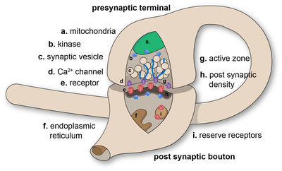
At the synapse, the plasma membrane of the signal-passing neuron (the presynaptic neuron) comes into close apposition with the membrane of the target (postsynaptic) cell. Both the presynaptic and postsynaptic sites contain extensive arrays of molecular machinery that link the two membranes together and carry out the signaling process. In many synapses, the presynaptic part is located on the terminals of axons and the postsynaptic part is located on a dendrite or soma. Astrocytes also exchange information with the synaptic neurons, responding to synaptic activity and, in turn, regulating neurotransmission.[10] Synapses (at least chemical synapses) are stabilized in position by synaptic adhesion molecules (SAMs)[1] projecting from both the pre- and post-synaptic neuron and sticking together where they overlap; SAMs may also assist in the generation and functioning of synapses.[12] Moreover, SAMs coordinate the formation of synapses, with various types working together to achieve the remarkable specificity of synapses.[11][13] In essence, SAMs function in both excitatory and inhibitory synapses, likely serving as the mediator for signal transmission.[11]
- ^ Foster M, Sherrington CS (1897). Textbook of Physiology. Vol. 3 (7th ed.). London: Macmillan. p. 929. ISBN 978-1-4325-1085-5.
- ^ Bennett MV (1966). "PHYSIOLOGY OF ELECTROTONIC JUNCTIONS*". Annals of the New York Academy of Sciences. 137 (2): 509–539. doi:10.1111/j.1749-6632.1966.tb50178.x. ISSN 0077-8923.
- ^ Kandel ER, ed. (2013). Principles of neural science (5th ed.). New York: McGraw-Hill. ISBN 978-0-07-139011-8.
- ^ Purves D, Williams SM, eds. (2004). Neuroscience (3rd ed.). Sunderland, Mass: Sinauer Associates. ISBN 978-0-87893-725-7.
- ^ Bennett MV, Zukin R (2004). "Electrical Coupling and Neuronal Synchronization in the Mammalian Brain". Neuron. 41 (4): 495–511. doi:10.1016/s0896-6273(04)00043-1. ISSN 0896-6273.
- ^ Makarenko V, Llinás R (1998-12-22). "Experimentally determined chaotic phase synchronization in a neuronal system". Proceedings of the National Academy of Sciences. 95 (26): 15747–15752. doi:10.1073/pnas.95.26.15747. ISSN 0027-8424. PMC 28115. PMID 9861041.
- ^ Korn H, Faure P (2003). "Is there chaos in the brain? II. Experimental evidence and related models". Comptes Rendus. Biologies (in French). 326 (9): 787–840. doi:10.1016/j.crvi.2003.09.011. ISSN 1768-3238.
- ^ Connors BW, Long MA (2004-07-21). "ELECTRICAL SYNAPSES IN THE MAMMALIAN BRAIN". Annual Review of Neuroscience. 27 (1): 393–418. doi:10.1146/annurev.neuro.26.041002.131128. ISSN 0147-006X.
- ^ Apodaca RL (2006-09-08). "Chemical Reviews on Wikipedia". doi.org. Retrieved 2025-02-02.
- ^ a b Perea G, Navarrete M, Araque A (August 2009). "Tripartite synapses: astrocytes process and control synaptic information". Trends in Neurosciences. 32 (8). Cell Press: 421–431. doi:10.1016/j.tins.2009.05.001. PMID 19615761. S2CID 16355401.
- ^ a b c Südhof TC (July 2021). "The cell biology of synapse formation". The Journal of Cell Biology. 220 (7): e202103052. doi:10.1083/jcb.202103052. PMC 8186004. PMID 34086051.
- ^ Missler M, Südhof TC, Biederer T (April 2012). "Synaptic cell adhesion". Cold Spring Harbor Perspectives in Biology. 4 (4): a005694. doi:10.1101/cshperspect.a005694. PMC 3312681. PMID 22278667.
- ^ Hale WD, Südhof TC, Huganir RL (January 2023). "Engineered adhesion molecules drive synapse organization". Proceedings of the National Academy of Sciences of the United States of America. 120 (3): e2215905120. Bibcode:2023PNAS..12015905H. doi:10.1073/pnas.2215905120. PMC 9934208. PMID 36638214.
Previous Page Next Page


