Our website is made possible by displaying online advertisements to our visitors.
Please consider supporting us by disabling your ad blocker.
Trigeminal nerve
This article includes a list of general references, but it lacks sufficient corresponding inline citations. (October 2014) |
| Trigeminal nerve | |
|---|---|
 Schematic illustration of the trigeminal nerve and the organs (or structures) it supplies | |
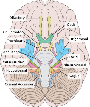
 Inferior view of the human brain, with cranial nerves labelled | |
| Details | |
| To | Ophthalmic nerve Maxillary nerve Mandibular nerve |
| Innervates | Motor: Muscles of mastication, tensor tympani, tensor veli palatini, mylohyoid, anterior belly of the digastric Sensory: Face, mouth, temporomandibular joint |
| Identifiers | |
| Latin | nervus trigeminus |
| MeSH | D014276 |
| NeuroNames | 549 |
| TA98 | A14.2.01.012 |
| TA2 | 6192 |
| FMA | 50866 |
| Anatomical terms of neuroanatomy | |
| Cranial nerves |
|---|
|
In neuroanatomy, the trigeminal nerve (lit. triplet nerve), also known as the fifth cranial nerve, cranial nerve V, or simply CN V, is a cranial nerve responsible for sensation in the face and motor functions such as biting and chewing; it is the most complex of the cranial nerves. Its name (trigeminal, from Latin tri- 'three' and -geminus 'twin'[1]) derives from each of the two nerves (one on each side of the pons) having three major branches: the ophthalmic nerve (V1), the maxillary nerve (V2), and the mandibular nerve (V3). The ophthalmic and maxillary nerves are purely sensory, whereas the mandibular nerve supplies motor as well as sensory (or "cutaneous") functions.[2] Adding to the complexity of this nerve is that autonomic nerve fibers as well as special sensory fibers (taste) are contained within it.
The motor division of the trigeminal nerve derives from the basal plate of the embryonic pons, and the sensory division originates in the cranial neural crest. Sensory information from the face and body is processed by parallel pathways in the central nervous system.
- ^ American Heritage Dictionary, 1969.
- ^ Pazhaniappan N (15 August 2020). "The Trigeminal Nerve (CN V)". TeachMeAnatomy. Retrieved 5 April 2021.
Previous Page Next Page


