Back خلية مخروطية Arabic Kolbacıqlar AZ Čepićasta ćelija BS Con (cèl·lula) Catalan خانەی قووچەکی CKB Čípek (oko) Czech Tap (synet) Danish Zapfen (Auge) German Κωνία Greek Konuseto EO
| Cone cells | |
|---|---|
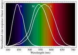
 Normalized responsivity spectra of human cone cells, S, M, and L types | |
| Details | |
| Location | Retina of vertebrates |
| Function | Color vision |
| Identifiers | |
| MeSH | D017949 |
| NeuroLex ID | sao1103104164 |
| TH | H3.11.08.3.01046 |
| FMA | 67748 |
| Anatomical terms of neuroanatomy | |
Cone cells or cones are photoreceptor cells in the retinas of vertebrates' eyes. They respond differently to light of different wavelengths, and the combination of their responses is responsible for color vision. Cones function best in relatively bright light, called the photopic region, as opposed to rod cells, which work better in dim light, or the scotopic region. Cone cells are densely packed in the fovea centralis, a 0.3 mm diameter rod-free area with very thin, densely packed cones which quickly reduce in number towards the periphery of the retina. Conversely, they are absent from the optic disc, contributing to the blind spot. There are about six to seven million cones in a human eye (vs ~92 million rods), with the highest concentration being towards the macula.[1]
Cones are less sensitive to light than the rod cells in the retina (which support vision at low light levels), but allow the perception of color. They are also able to perceive finer detail and more rapid changes in images because their response times to stimuli are faster than those of rods.[2] Cones are normally one of three types: S-cones, M-cones and L-cones. Each type expresses a different opsin: OPN1SW, OPN1MW, and OPN1LW, respectively. These cones are sensitive to visible wavelengths of light that correspond to short-wavelength, medium-wavelength and longer-wavelength light respectively.[3] Because humans usually have three kinds of cones with different photopsins, which have different response curves and thus respond to variation in color in different ways, humans have trichromatic vision. Being color blind can change this, and there have been some verified reports of people with four types of cones, giving them tetrachromatic vision.[4][5][6] The three pigments responsible for detecting light have been shown to vary in their exact chemical composition due to genetic mutation; different individuals will have cones with different color sensitivity.
- ^ "The Rods and Cones of the Human Eye". HyperPhysics Concepts - Georgia State University.
- ^ Kandel, E.R.; Schwartz, J.H; Jessell, T. M. (2000). Principles of Neural Science (4th ed.). New York: McGraw-Hill. pp. 507–513. ISBN 9780838577011.
- ^ Schacter, Gilbert, Wegner, "Psychology", New York: Worth Publishers,2009.
- ^ Jameson, K. A.; Highnote, S. M. & Wasserman, L. M. (2001). "Richer color experience in observers with multiple photopigment opsin genes" (PDF). Psychonomic Bulletin and Review. 8 (2): 244–261. doi:10.3758/BF03196159. PMID 11495112. S2CID 2389566.
- ^ "You won't believe your eyes: The mysteries of sight revealed". The Independent. 7 March 2007. Archived from the original on 6 July 2008. Retrieved 22 August 2009.
Equipped with four receptors instead of three, Mrs M - an English social worker, and the first known human "tetrachromat" - sees rare subtleties of colour.
- ^ Mark Roth (September 13, 2006). "Some women may see 100,000,000 colors, thanks to their genes". Pittsburgh Post-Gazette. Archived from the original on November 8, 2006. Retrieved August 22, 2009.
A tetrachromat is a woman who can see four distinct ranges of color, instead of the three that most of us live with.