Back Heiligbeen AF عجز (تشريح) Arabic Oma sümüyü AZ Һигеҙгүҙ һөйәк BA Askorn kroazell BR Krstačna kost BS Sacre Catalan سێبەندە CKB Křížová kost Czech Ыса CV
| Sacrum | |
|---|---|
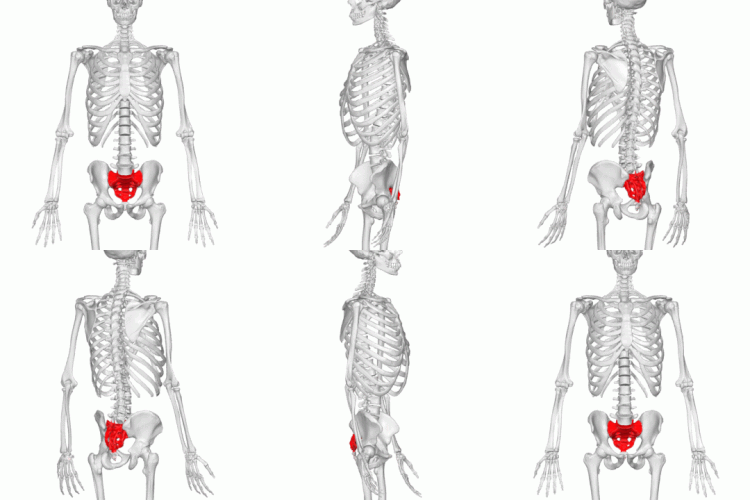
 Position of the sacrum in the pelvis | |
 Animation of the sacrum in the human skeleton | |
| Details | |
| Pronunciation | (/ˈsækrəm/ or /ˈseɪkrəm/) |
| Location | Base of the vertebral column |
| Identifiers | |
| Latin | os sacrum |
| MeSH | D012447 |
| TA98 | A02.2.05.001 |
| TA2 | 1071 |
| FMA | 16202 |
| Anatomical terms of bone | |
The sacrum (pl.: sacra or sacrums[1]), in human anatomy, is a large, triangular bone at the base of the spine that forms by the fusing of the sacral vertebrae (S1–S5) between ages 18 and 30.[2]
The sacrum situates at the upper, back part of the pelvic cavity, between the two wings of the pelvis. It forms joints with four other bones. The two projections at the sides of the sacrum are called the alae (wings), and articulate with the ilium at the L-shaped sacroiliac joints. The upper part of the sacrum connects with the last lumbar vertebra (L5), and its lower part with the coccyx (tailbone) via the sacral and coccygeal cornua.
The sacrum has three different surfaces which are shaped to accommodate surrounding pelvic structures. Overall, it is concave (curved upon itself). The base of the sacrum, the broadest and uppermost part, is tilted forward as the sacral promontory internally. The central part is curved outward toward the posterior, allowing greater room for the pelvic cavity.
In all other quadrupedal vertebrates, the pelvic vertebrae undergo a similar developmental process to form a sacrum in the adult, even while the bony tail (caudal) vertebrae remain unfused. The number of sacral vertebrae varies slightly. For instance, the S1–S5 vertebrae of a horse will fuse, the S1–S3 of a dog will fuse, and four pelvic vertebrae of a rat will fuse between the lumbar and the caudal vertebrae of its tail.[3]
The Stegosaurus dinosaur had a greatly enlarged neural canal in the sacrum, characterized as a "posterior brain case".[4]
- ^ Oxford Dictionaries and Webster's New College Dictionary (2010) admit the plural sacrums alongside sacra; The American Heritage Dictionary, Collins Dictionary and Webster's Revised Unabridged Dictionary (1913) give sacra as the only plural.
- ^
Kilincer, Cumhur; et al. (2009). "Sacrum anatomy". Scientific spine. Trakya Üniversitesi Rektörlüğü, Yerleşkesi, 22030 Edirne, Turkey: Self. Retrieved 8 November 2015.
{{cite web}}: CS1 maint: location (link) - ^ "Skeletal system" (PDF). Dept. of Biology. Gambier, Ohio: Kenyon College. Retrieved 9 November 2015.
- ^ Galton, P.M.; Upchurch, P. (2004). "Stegosauria". In Weishampel, D.B.; Dodson, P.; Osmólska, H. (eds.). The Dinosauria (2nd ed.). University of California Press. p. 361. ISBN 0-520-24209-2.